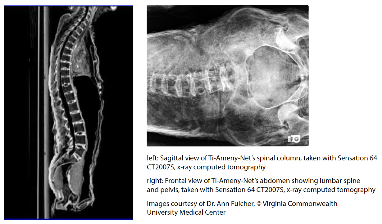
X-rays and more than 25,000 CT-scans of Ti Ameny Net’s body were taken in 2010 by Dr. Ann Fulcher, Professor and Chair, Department of Radiology, Virginia Commonwealth University Medical Center, and a 1983 alumna of the University of Richmond, working with a current University of Richmond student, Caroline Cobert, ’12.
The image on the left shows the torso from the side with the entire spinal column visible and in alignment. The diaphragm is still preserved in the chest. Packing material, including linen, resin, and an unidentified material with a honeycombed structure, possibly packs of natron, can be seen in the pelvis, at the bottom of the image.
The x-ray image on the right shows developmental scoliosis (curvature of the lower spine), which was untreated and caused inflammatory growth of her vertebrae. The absence of scarring on the pelvis bones indicates she did not give birth to any children.

Up next: Computer Tomography – CT Scans
