So long, Nate!

This summer Trawick lab bids farewell to Nate Lawrence, who graduated in the spring but stayed on to continue working on his research project over this summer.  Nate starts graduate school in electrical engineering at Boston University this fall.

Nate is a great example of a student who took advantage of the opportunity to do research here.  He started working with me as a first year student, learning to use the atomic force microscope in our lab.  His research continued here over the summers, supported by a summer fellowship from the Virginia Foundation for Independent Colleges, and later by a grant from the Research Corporation.  Over the course of four years, he has become a highly skilled researcher and a real expert in the field, and his work was presented at several national physics conferences.  During his four years at UR, Nate spent one summer doing research at Boston University, and also spent a semester abroad in Scotland.  He won the Robert Edward Loving Award in Physics for promise for advanced study in physics.

Nate’s expertise will be sorely missed around here.  So long, Nate, and stay in touch!

Yee-ouch, them pins is pointy! (Our highest resolution microscope image ever)

Over the last few months, we’ve been collaborating with Mike Leopold and his students in the chemistry department to use our atomic force microscope to image their nanoparticle films.  We just purchased a small number of extra sharp (and extra expensive) tips for the microscope that allow us to resolve surface features about 1 nanometer in size.  This image of a thin gold film that was coated over a mica surface is our first picture with the new tips.  The mica is mostly very smooth, except for the big crevasse in the lower right hand corner.  The surface roughness that you see is from the gold film, which clumps together to form small “grains” about 20-30 atoms across, or about 10 nanometers.

baregold-0008.png

Take a moment to appreciate the scale of this picture.  The vertical scale at the right shows that the darkest areas are about 5 nanometers lower than the whitest areas; most of the surface roughness is on the order of about two nanometers.  The entire image is 500 nanometers across, or half a micron.  If it shows up on your computer screen as 4 inches across, then the width of a human hair on the same scale would be about fifty feet! 

This image was taken by my student, Nate Lawrence.

Matt needs more research students!

One of the nice things about teaching is seeing my students graduate and go on to do cool things elsewhere.  Of course, it’s not so nice when my entire lab group graduates all at once, as happened this spring!  (Nate…Jill…Brian…David!  Please come back!)

Anyway, I just want to put the word out there that I’m very actively looking for some students to join me in fall of 2008 and beyond.  I have a handful of projects I’ll be involved in; some projects will entail using the cool and expensive atomic force microscope in our lab; others will involve more computer coding and analysis.  I’m especially interested in finding a strong first or second year student.  I’m imagining working for course credit initially,  but I also have some grant money that could support a student over the summer.

One project I’ve written about here recently involves writing software to correct for distortion in atomic force microscope images.  This is a neat project involving a real-life application of numerical computing and image analysis; the resulting software package will be shared with potentially hundreds of scientists and engineers using around the world who use scanning probe microscopes.  Requirements: must be comfortable programming in C/C++, and must know some calculus.  (Everything else is learn-as-you-go.) 

Please contact me if you or anybody you know might be interested!

New results: distortion removed from AFM images

I thought I’d share a nifty result out of the lab that involves correcting distortion in the images we take with our atomic force microscope (AFM).

An AFM works by physically positioning a very sharp tip over a sample and rastering it back and forth over the image.  To look at nanometer-scale features as we do in our lab, this means having to physically position the tip over the surface with nanometer precision.  It turns out that even the best AFMs in the world run into a problem called thermal drift.  When the temperature in the room changes–by even a tenth of a degree–some parts of the microscope or the sample are always going to expand or contract with temperature by just a little bit more than other parts.  And that means that the tip can slowly move from where you thought it was.   Imagine scanning an image slowly–over several minutes–while the thing you’re imaging is moving under you.  The result is an image that can appear stretched, compressed, or badly skewed.

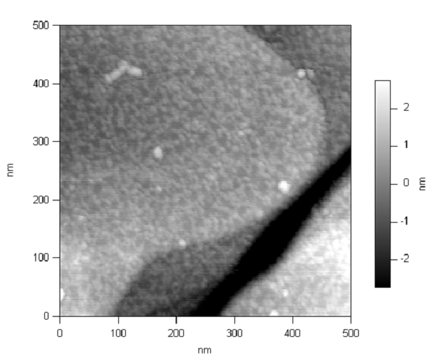
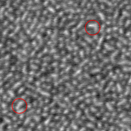
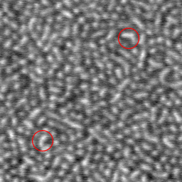
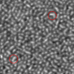
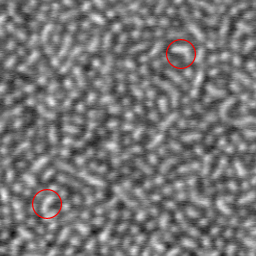
Check two pictures below, which are portions of two scans that were taken right after each other.   (One was scanned going up, the other scanned going down.)  The circled feature in the upper right is in the same place in each image, but the circled feature in the lower left is shifted slightly.  That’s because both images are slightly distorted from thermal drift.  On the left image, the distance between the two features appears to be is 400 nanometers.  On the right, it appears to be 357 nanometers.

complete_set_small2_i3.jpg   complete_set_small2_i4.jpg

Below are the same two images, after each one has been corrected using our new technique.  (They were corrected independently of each other; if all we did was use one as a guide for the other, that would be cheating!)  Now, in both corrected images, the features appear to be the same distance apart: 382 nanometers.

complete_set_small2_i1.jpg   complete_set_small2_i2.jpg

The way we corrected each image was by rescanning a small sliver from down the center of each image.  The rescanned sliver acts as a “key” to remove the distortion from the main image.  There’s actually a lot of computation involved in it, all performed by some numerically intensive computer software written by me and my student, Brian Salmons.

We still have a bit of testing and refining to do on our technique, but we hope we’ll have some published results soon.  (Watch for it in a journal near you!)  Of course, we’ll make our code available to the public.

 Later on, there are several features and enhancements I’d like to add, and I’m actively looking for a physics or computer science student at Richmond to work with.  If you’re interested in working on this project with me, let me know.

A nice picture of our microscope

Here are Jill, Nate, and me looking at some data on the our atomic force microscope (AFM).  The microscope is in the background inside the enclosure.  In the foreground is a rack of electronics that Cosmin and I are using to modify its feedback response mechanism.  I’m always eager to meet students who may be interested in working with me in my lab.  If you’re a young, potential physics major, or even a prospective student, please contact me. 

the-microscope.jpg�