How Do You Separate Imagination From Reality?
Have you ever thought of how your brain separates imagination from reality? If we can imagine movies we have just seen or the faces of people we just met, then what separates the actual visual perception of these experiences from our visual mental imagery? Sometimes it can be difficult to separate these two processes, like the man in this video here, who is using the Oculus Rift with a roller coaster video game. Skip to the 1:20 mark to see his reaction.

In this video, the man testing the Oculus Rift freaks out when he goes over the edge of the roller coaster. Here, the video game is so immersive that he actually feels like he is experiencing everything on the screen. Look how he leans back and tenses up as he approaches the top of the roller coaster. So how does our brain deal with reality as we see it (visual perception), and visual imagery (imagination)?
Visual Perception and Visual Imagery Explained
Visual perception is the intake of sensory information through the eyes and the distribution of this information through the optic nerves, and across the optic chiasm as it makes its way to the primary visual cortex in the back of the brain. More information about visual perception can be found here.
On the other hand, visual imagery is a top-down process, meaning that knowledge is retrieved from different parts of the brain in order to construct a certain image or scene.
Studies About Visual Perception and Visual Imagery
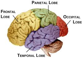
A study by Giorgio Ganis with the help of Harvard University and Massachusetts General Hospital, revealed that there is some overlap between visual perception and visual imagery especially in the frontal and parietal cortices. This study also found that “the regions engaged by visual imagery across the brain were a subset of those engaged during visual perception”. So, visual imagery uses some of the same areas of the brain as visual perception, but visual perception still utilizes other areas, such as the optic pathways used to take in sensory information from the eyes.
However, scientists at the University of Wisconsin-Madison published a study in 2014, which revealed some interesting new findings. Although visual imagery and visual perception use the same regions of the brain, the neural impulses actually flow in different directions in the parieto-occipital cortices, which further separates these two processes.

One of the many functions of the parietal lobe is to process and integrate sensory information such as touch, while the occipital lobe is responsible for processing visual information from the eyes.
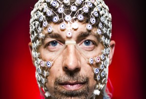
Dr. Barry Van Veen and his team at The University of Wisconsin-Madison measured cortical interactions among different parts of the brain when patients viewed a short, 1 minute, movie, and then tried to replay the film in their head with as much detail as possible. Participants did this with their eyes open, and again with their eyes closed. They were also asked to imagine themselves riding a magical bike to account for daydreaming and free imagery.

The team used high-density electroencephalography (hdEEG), which combines excellent temporal resolution of EEG recordings with high spatial resolution and quantified the results with the Granger causality (GC) and dynamic causal modeling (DCM). Using these methods, the scientists were able to estimate cortical activity directionality. These methods were key for finding the reversal of the neural signal flow, especially along the visual dorsal stream for visual imagery and perception.
These results are consistent with other literature of the time as a study by Andrea Mechelli, showed that visual perception is a bottom up process that flows from the occipital lobe to the temporo-occipital cortex while visual imagery is a top-down process that moves from the prefrontal cortex and then to the previously stated cortices in the brain.
Time magazine published an article about a study with a group of researchers at Cambridge university, which stated that some people may have a more difficult time differentiating reality from the imaginary. The paracingulate sulcus is part of the prefrontal cortex that develops late in pregnancy and it is missing in 27% of people. Participants who lacked the paracingulate sulcus performed significantly worse when trying to remember words they had read and when prompted with words that they had not been tested with, claimed that they had read them with confidence. These participants were also unaware of their inaccurate memories.

Representations of reality vs imagination in popular culture
The idea of questioning reality has been incorporated into many different TV shows and movies such as Wilfred, The Matrix, and most recently, Inception.
In the film, the characters can enter a dream like state, in which they can forge their own realities. This experience makes it very difficult to differentiate what is real and what is not. I highly recommend watching this movie if you want to have your mind blown.
The ground breaking research done at The University of Wisconsin-Madison involving the reversal of neural impulses in the brain during visual perception and visual imagery will hopefully lead to more studies done involving the two processes. This work also paves the way for more studies about the way visual imagery and visual perception work during dreaming. So what is real? Grab some popcorn, watch inception, and start to question the world around you.
References
http://www.sciencedirect.com/science/article/pii/S1053811914004662
http://cercor.oxfordjournals.org/content/14/11/1256.full.pdf+html
http://www.ncbi.nlm.nih.gov/pubmed/22593753-source