Can You or Anyone Else Acquire Sociopathy? Yes. Read How!

Do you have commitment issues? Do many of your friends and peers consider you irresponsible or immature for your age? Are you impulsive? If you have answered yes to all of these questions you might be a sociopath…but hey, at least you aren’t a psychopath!

What is sociopathy?

“Sociopathy is a personality disorder associated with irresponsible and unreliable behavior that is not personality advantageous; an inability to form lasting commitments or relationships; egocentric thinking; and a marked degree of impulsivity.”

— (Ward, 2015)

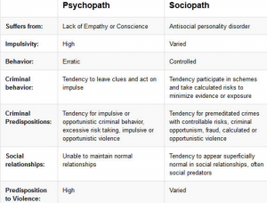
Psychopath verses sociopath

sociopath

The hyenas from the classic film “Lion King” are Disney characters that display psychopathic tendencies

Hyenas

Scar from the classic film “Lion King” is a Disney character that is an exemplary illustration of a sociopath

Scar

How do people become sociopaths/psychopaths?

Research indicates that specific genes predispose people to psychopathy and sociopathy; however, these genes are usually dormant.  The chances of these predisposed genes actually being expressed greatly increases in the presence of environmental triggers. However, genes are not always necessary to “turn” a person into a sociopath. Often times, an abusive or rough childhood or a traumatic head injury is all that is needed (Bree, et al., 1998). Once in a while though, a serious head injury to a specific part of the brain is all that is needed to turn an average, well-functioning member of society into an out of control, impulsive sociopath with no regards for others.

Acquired sociopathy refers to a person who was “normal” that, after a traumatic brain injury, “turned” into a sociopath.

Cases of acquired sociopathy

Phineas Gage’s Accident

The most well-known case of acquired sociopathy is that of Phineas Gage. Before Phineas Gage had a metal rod pass through his left cheek and out the top of his head, he was denoted as a serious, industrious, and energetic person who was responsible and smart. Yet after the incident, his peers claimed that he was childish, irresponsible, and thoughtless of others. This was a drastic change of personality. Although Gage was considered lucky to be alive, the metal rod damaged his left orbitofrontal/ventromedial region and the left anterior region. Severe trauma to specific regions of the brain can cause a person to undergo marked personality changes, as seen with Phineas Gage (Damasio, 1994). Another example of a similar case is the J.S case in 1996. This incident also supports the idea that brain trauma alone can lead to acquired sociopathic behaviors.

J.S. is a 56-year-old man who worked as an electrical engineer. In November 1996, he was found collapsed and unconscious with evidence of trauma to the right frontal region. It was noted that his behavior was abnormal. He behaved bizarrely; riding around on a hospital gurney, being uncooperative, and having unpredictable episodes of aggression such as when he assaulted and wounded a staff member, threw objects and furniture at people, and behaved aggressively towards other patients. However he had no psychiatric history prior to his accident and before the incident, he was described by a relative as being a quiet, rather withdrawn person who was never aggressive (Blair, 2000).

Behind the scenes of executive functioning

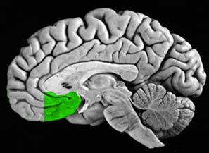
The ventromedial prefrontal cortex (vmPFC) plays an important role in emotional reactions. This region communicates with the dorsomedial thalamus, temporal cortex, ventral tegmental area, olfactory system, amygdala, cingulate pam1
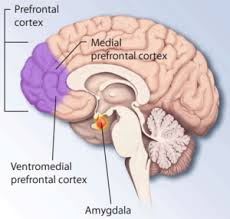
cortex, lateral hypothalamus, and other regions of the frontal cortex, including the dorsolateral prefrontal cortex. People with ventromedial prefrontal lesions show impulsive behavior and often display outbursts of inappropriate anger. They are able to explain the implications of complex social situations but often respond inappropriately when they find themselves in these situations. Evidence indicates that the ventromedial prefrontal cortex is involved in making moral judgments (Ward, 2015) (Damasio, et al., 1994).

In summation

Neuroscientists have identified areas of the brain related to sociopathic behaviors. Subtle damage to the amygdala, a brain region that helps us process our emotions, may explain why sociopaths cannot read or express emotions properly. Sociopathic behaviors are also associated with injury to the ventromedial region of both frontal lobes. This is where the umbrella term  for the management of cognitive processes, executive functioning, comes into play. Executive function is associated with the prefrontal cortex (PFC) which is what tends to be damaged in patients with acquired sociopathy.

References

Bree, M. B., Svikis, D. S., & Pickens, R. W. (1998). Genetic influences in antisocial personality and drug use disorders. Drug and Alcohol Dependence,49(3), 177-187. doi:10.1016/s0376-8716(98)00012-x

Blair, R. J. (2000). Impaired social response reversal: A case of `acquired sociopathy’ Brain,123(6), 1122-1141. doi:10.1093/brain/123.6.1122

Carlson, N. R. (2012). Emotion. In Physiology of behavior (11th ed., pp. 360-376). Pearson.

Damasio, A. R. (1994). Descartes’ error: Emotion, reason, and the human brain. New York: Putnam.

Damasio, H., Grabowski, T., Frank, R., Galaburda, A., & Damasio, A. (1994). The return of Phineas Gage: Clues about the brain from the skull of a famous patient. Science,264(5162), 1102-1105. doi:10.1126/science.8178168

Ward, J. (2015). The executive brain. In The student’s guide to cognitive neuroscience (3rd ed., pp. 345-371). Psychology Press.

The Bilingual Brain: Twice as smart?

All of us have experienced the difficulties of learning a second language. Whether your reason for struggling through a whole new set of grammar and vocabulary was purely to satisfy your COM2 requirement, or whether you had a true personal interest in learning another language or were raised in a bilingual household, most of us can agree that there are certain benefits to bilingualism.

b4

But did you ever think that bilingualism could actually be benefiting your brain?

Research shows that bilingualism does just that. “How could it possibly help my brain just to know another language?” you might ask. Here’s the basic idea:

In a 6-week study at Penn State, researchers scanned the brains of American, English-speaking subjects before and after they learned Chinese vocabulary. The brain scans after the 6 weeks ended showed some interesting findings:

  1. Those who were successful at learning the new language showed increased connectivity within their brains
  2. Their brain networks were better integrated, leading to increased flexibility and efficiency
  3. Their brains showed increased density in grey matter, as well as strengthened white matter

The study showed that these findings were consistent across the average life span, meaning that even older brains showed these same structural and functional b5changes with language learning. The short span of the study, only 6 weeks, also
showed that these changes could occur rapidly with shorter, intensive periods of language learning.

The brain itself clearly changes due to the presence of bilingualism, but there are other benefits to it as well:

  • A study done by Dr Thomas Bak found that, on average, multilingual people develop dementia four years later than monolinguals
  • Bak also found that multilingual young adults had better attention spans and could more easily ignore unnecessary information

b1So why doesn’t everyone just learn every language known to man? Well, there are some difficulties to bilingualism as well.

b3

That moment when you start thinking in two languages at the same time

For instance, it was found through an fMRI study that native and secondary languages are separated from one another in Broca’s area, but are not separated in Wernicke’s area. This finding illustrates that often in learning a foreign language it isn’t comprehension (controlled by Wernicke’s area) that is difficult but instead is the actual formation of the words into speech (controlled by Broca’s area) that causes problems.

Interestingly, it is just those foreign phonemes, or bits of sound that make up words, that adults struggle with that their infants are babbling to them on a daily basis. Babies’ brains are equipped to learn whatever language is taught to them, and so they can access phonemes of foreign languages before their own language’s phonemes take over fully. It is this concept that makes it harder for adults to access the phonemes that make up foreign languages and is often why adult speakers never develop a perfect accent in a second or third language.

Hopefully by now you’re convinced; learning a second language has numerous benefits not only to your brain structure but also to your life. Here’s a short summary of some of the benefits of bilingualism:

The Back Of Your Hand Changes And So Do Your Memories

Crime and Memory 

Eyewitness testimony is paramount in determining the outcome of judicial proceedings. This makes sense considering how much we rely on our memory on a day-to-day basis. However, we’ve all forgotten where we parked the car, and we’ve all found ourselves walking into a room without remembering what our intentions were. While these are minor slip-ups, which we can write off, some of us may know what it’s like to be accused of saying or doing something we don’t believe we said or did. This might cause a bit of tension between or in groups of people, but being accused and convicted of a crime we did not commit is something that very few people have experienced.

According to Innocent Project – a non-profit organization that uses DNA testing to help exonerate wrongly convicted criminals – over 70% of 239 convictions based on eyewitness testimony have been overturned using genetic testing. (1) This makes us question why we use eyewitness testimony at all, and more importantly, makes us think about the accuracy of our own memories.

List of people exonerated using DNA testing

Click here if you would like to see part 2 of this documentary or here if you would like to view another documentary regarding crime and memory.

How the Brain Lies

As medical advancements and psychological research continues to progress, society (particularly the judicial system) is obligated to keep up, and we the people will eventually be left wondering: can you read my mind? While technology hasn’t progressed to mind-reading levels, scientists have made a few discoveries regarding the brain and what it looks like when someone is lying, even if they truly believe they are telling the truth.  Researchers have discovered two different brain regions that may respond differently to true and false recognition. Apparently, our medial temporal lobe (MTL): parahippocampal gyrus shows greater activity during true recognition than false recognition. However, our hippocampus reacts equally to both. (2) Similar results have also been shown in other regions of the brain including the bilateral occipital cortices and the right parahippocampal gyrus. (3)

With that being said, science is still too far away to implement brain scanning lie detectors, and judges are far from accepting fMRIs as legitimate evidence for a crime. Hence, it’s at least safe to say that we can’t exactly read anyone’s mind yet. But if technology could help us detect our own lies, then one has to wonder what he or she would want to know or find out, if anything.

Why the Brain Lies

Although we get mad at ourselves for misplacing our keys, forgetting may be an adaptation rather than a design fault, and it may be a design fault that we actually want to have. After all, we don’t want or care to know where we parked the car last week. We need to know where we parked it today, and we don’t want to have to sift through all of our memories to do so. It’s efficient to have prioritized memories. However, when we forget we often question how or why this happens. From a cognitive point of view, neuroscientists tend to group causes of forgetting into three categories involving encoding, storage, and retrieval. Within each category there are many hypotheses that explain how forgetting can occur. Maybe we’re not in the correct setting to remember something, or perhaps similar memories are overlapping and are hard to tell apart. Whatever the case, the fact that our brain can drop the ball at so many levels should tell us something about the power of forgetting. In fact, sometimes people don’t want to know a particular piece of information, or they intentionally block out memories. (4)

Furthermore, even if forgetting is unintentional, and we fully believe in the accuracy of our memories, then why has criminology proven us wrong, and why do we tell different versions of the same story? If humans can make eyewitness errors regarding the most horrendous crimes and forget where their phones are, then who are we to not question everything in between? Humans do use past experiences to judge how the future will turn out, but if our past experiences can only be so accurate, then how do we know we’re making the right choices going forward? Looking back, it’s interesting to think about what we do and do not remember. However, the more important question is if we would want to remember everything when we do reminisce.


References

(1) Arkowitz, H., & Lilienfeld, S. O. (2009, January 8). Why Science Tells Us Not to Rely on Eyewitness Accounts. Retrieved from                                                   http://www.scientificamerican.com/article/do-the-eyes-have-it

(2) Cabeza, R., Rao, S. M., Wagner, A. D., Mayer, A. R., & Schacter, D. L. (2001). Can medial temporal lobe regions distinguish true from false? An event-related functional MRI study of veridical and illusory recognition memory. Proceedings of the National Academy of Sciences, 98(8), 4805-4810. doi:10.1073/pnas.081082698

(3) Okado, Y., & Stark, C. (2003). Neural processing associated with true and false memory retrieval. Cognitive, Affective, & Behavioral Neuroscience, 3(4), 323-334. doi:10.3758/cabn.3.4.323

(4) Ward, J. (2015). The remembering brain. In The student’s guide to cognitive neuroscience (3rd ed., pp. 195-230). Hove, East Sussex: Psychology Press.

Understanding Attention Deficit Dis– Oh look, Squirrel!

Take a look at the following pictures and cartoons…

Did you laugh? Chances are you’ve probably seen these types of pictures before, or heard jokes about attention disorders. While these jokes are funny, there is little that’s very funny about this disorder. 


Background About the Disorder:

Attention disorders are extremely common among children. There are no distinct physical signs; the disorder is identified through characteristic patterns of behavior. There are three primary types attention disorders that fall under the umbrella of Attention Deficit Hyperactive Disorder: hyperactive (known as ADHD), deficit (known as ADD) and a combination between the two. Combined presentations is the most common subtype of ADHD.

ADHD is one of the most common childhood disorders and can continue throughout adolescence into adulthood. ADHD is more common among very young children prior to entering school, while ADD is often not diagnosed until middle school. There is thought to be a genetic basis for this disorder, and it is influenced by hereditary factors. Boys are four times as likely as girls to have this disorder, and 11% of American children from age 13 to 18 have this disorder. (1) There is estimated to be 6.4 million children in the US diagnosed with ADHD. Most children “outgrow” this disorder, but some carry their difficulties into adulthood. In fact, approximately 10 million Americans are diagnosed with ADHD. (8) Of the children referred to mental health clinics, 50% are children with ADHD-related problems.


Possible developmental pathway for ADHD. Retrieved from Google.


There are distinct differences in symptoms between ADD and ADHD. To diagnose a child, however, there are certain criteria that must be met. The symptoms must be present before age 12 and in two or more settings (home, school, work, peer relationships).

Symptoms of Attention Deficit Disorder 
–  Be easily distracted, miss details, forget things, and frequently switch from one activity to another
–  Become bored with a task after only a few minutes, unless they are doing something enjoyable
–  Have difficulty focusing attention on organizing and completing a task or learning something new
–  Have trouble completing or turning in homework assignments, often losing things
–  Not seem to listen when spoken to
–  Daydream, become easily confused, and move slowly
–  Have difficulty processing information as quickly and accurately as others
–  Struggle to follow instructions.
Symptoms of Attention
Deficit Hyperactive Disorder
–  Fidget and squirm in their seats
–  Talk nonstop
–  Dash around, touching or playing with anything and everything in sight
–  Have trouble sitting still during dinner, school, and story time
–  Be constantly in motion
–  Have difficulty doing quiet tasks or activities.


https://www.youtube.com/watch?list=PLEbm7wJ3WiYy45JHXcdEzsiE-RxGG4_Xe&t=5&v=rp1IleFD4D0

Happening in the Brain?

Brain imaging studies have revealed that, in youth with ADHD, the brain matures in normally but is delayed, on average, by about 3 years.(3) The delay is most pronounced in brain regions involved in thinking, attention, and planning. For example, there is often dysfunction and lower activity in the prefrontal lobe, which is involved in inhibition and executive function.

joc20194f2

Graph shows the caudate volume and the cerebellar volume of patients with ADHD and without ADHD. Notice how ADHD patients consistently have lower volumes. Retrieved from Castellanos, 2002.

More recent studies have found that the outermost layer of the brain, the cortex, shows delayed maturation overall,(4) and a brain structure important for proper communications between the two halves of the brain, the corpus callosum, shows an abnormal growth pattern.(5) Additionally, a study has shown that children with ADHD have smaller cerebellums. The caudate nucleus volume was found to be lower and asymmetrical in children with ADHD. The caudate nucleus is one of the three primary structures of the basal ganglia and is associated with voluntary movement, learning, memory, sleep, and social behavior.


Sagittal view of the posterior cingulate cortex (highlighted in green). Retrieved from Google.

Researchers use fMRI to study the brain of those with and without ADHD. They were specifically looking at the posterior cingulate cortex and the medial prefrontal cortex. The posterior cingulate cortex and the medial prefrontal cortex are central nodes in the default mode network (DMN) – a network of brain regions that are active when the individual is not focused on the outside world and the brain is at wakeful rest.

Sagittal view of the medial prefrontal cortex. Retrieved from Google.

Researchers found that in people without ADHD, the activity in those brain regions were synchronous, but in ADHD children and adults, it was no longer synchronized. A newer study found that adults who were diagnosed with ADHD as children have a restored normal synchrony pattern, and their brains look like those of people who never had ADHD. (8)


Resources:

(1)http://www.nimh.nih.gov/health/topics/attention-deficit-hyperactivity-disorder-adhd/index.shtml

(2)Konrad, Kerstin, and Simon B. Eickhoff. “Is the ADHD Brain Wired Differently? A Review on Structural and Functional Connectivity in Attention Deficit Hyperactivity Disorder.” Human Brain Mapping Hum. Brain Mapp. 31.6 (2010): 904-16. Web.

(3)Shaw P, Eckstrand K, Sharp W, Blumenthal J, Lerch JP, et al. Attention-deficit/hyperactivity disorder is characterized by a delay in cortical maturation. Proc Natl Acad Sci U S A. 2007 Dec 4;104(49):19649–54. Epub 2007 Nov 16. PubMed PMID: 18024590; PubMed Central PMCID: PMC2148343.

(4)Shaw P, Malek M, Watson B, Sharp W, Evans A, Greenstein D. Development of cortical surface area and gyrification in attention-deficit/hyperactivity disorder. Biol Psychiatry. 2012 Aug 1;72(3):191–7. Epub 2012 Mar 13. PMID: 22418014.

(5)Gilliam M, Stockman M, Malek M, Sharp W, Greenstein D, et al. Developmental trajectories of the corpus callosum in attention-deficit/hyperactivity disorder. Biol Psychiatry. 2011 May 1;69(9):839–46. Epub 2011 Jan 17. PMID: 21247556.

(6)Castellanos F, Lee PP, Sharp W, et al. Developmental Trajectories of Brain Volume Abnormalities in Children and Adolescents With Attention-Deficit/Hyperactivity Disorder. JAMA. 2002;288(14):1740-1748. doi:10.1001/jama.288.14.1740.

(7)https://en.wikipedia.org/wiki/Caudate_nucleus

(8)http://mcgovern.mit.edu/news/news/inside-the-adult-adhd-brain/

Decoding Visual Illusions: What You See is Not Always the Truth

big-eye

So seeing is believing right?  Think again…

The whole concept behind visual illusions is that our minds make assumptions about the sensory information we perceive.  A visual illusion occurs when our minds perceive an image that is different from the objective reality.  This may happen for a variety of reasons such as over stimulation of the brain and eyes or the brain and eyes making unconscious inferences.  The bottom line is that what we see isn’t always necessarily the truth…

This visual illusion is called the Hermann Grid. Notice how the black dots are coming and going and each individual dot will become white when you focus on it.

Let’s take a look at the Hermann Grid, a classic example of visual illusions that deals with contrast and colors.  Why are the black dots coming and going?  Why does any particular dot become white when you focus on it?

Visual sensory information is accepted by light receptive cones and rods.  The receptors pass the information down through intermediary cells, such as retinal ganglion cells.  However, here’s the twist: there are 125 million receptors in the retina and only one million retinal ganglion cells.  Therefore, before any information reaches the brain, all visual input is being compressed at a ratio of 125 to 1.  No wonder the “image” your light receptors receive is not the same “image” your brain receives.

Lateral Inhibition

The retinal ganglion gathers stimuli from a small area of light receptors.  Many ganglion cells then pass along the average of this stimuli.  However, lateral inhibition cells are a bit more complex.  For lateral inhibition cells, stimuli from the center of the receptive field is “positive” and stimuli from the edges of the receptive field is “negative.”  This becomes useful when the eyes are presented with edges because edges are where high contrast meet low contrast.  In the case of the Hermann Grid, the white dot in the middle is a “positive” stimulus but the surrounding white creates an even bigger “negative” stimulus.  Therefore, the middle white dot is perceived as white when we focus on it and the surrounding white lines are perceived as grey.  .

We can conclude from this that what we see may not be the truth.  Would it be too much of a stretch to take this lesson learned from visual illusions and apply it to our daily lives?  For example, if we catch a glimpse of our new crush talking to a girl or a guy we often jump to conclusions that he or she likes that person more than us.  But this may not be true, in fact often times that it is not the case.  As visual illusions teach us, seeing should not be believing.

Here is a list of optic illusions if you are interested in looking at a few more! Clicking on the name of the illusion will explain how the illusion works. Enjoy!

References:

Eustis, D. (n.d.). Contrast Effects. Retrieved September 25, 2015, from http://cs.brown.edu/~deus/courses/optical/Contrast1.htm
Lateral Inhibition. (2015). Retrieved September 25, 2015, from http://www.indiana.edu/~p1013447/dictionary/lat_i.htm

Left Brain vs. Right Brain- Distinguishing between Pseudoscience and Science.

What is Hemispheric Dominance?

We only have one brain, but the idea of being left or right brained has become popularized by TV shows, movies, online psychological tests, and even classroom settings where teaching styles vary based off brain hemispheric dominance. Moreover, many articles on the Internet are designed to help you self-diagnose hemispheric dominance such as the basic tests shown below.

Screen Shot 2015-09-05 at 9.54.33 PM

Folded hands test.

Motor function is controlled by the brain contralaterally. In other words, the left side of the brain controls the right side of the body and vice versa. Hence, if you place the left thumb above the right when clasping your hands, you are said to be right brained. Likewise, you are said to be left brained if you naturally place your right thumb above your left.
Screen Shot 2015-09-05 at 9.55.26 PM

If you sleep with the same person frequently, the side of the bed you sleep on indicates which side of your brain is dominant:

The idea of hemispheric dominance derives from the fact that the cerebral hemisphere is divided into right and left hemispheres. The two hemispheres are connected by the corpus callosum, a thick band of nerve fibers. Research says that each hemisphere of the brain is dominant for different tasks. For example, the left hemisphere is dominant for language, math, and logic, while the right hemisphere is dominant for spatial abilities, facial recognition, music, and visual imagery.

So do you think you’re left brained or right brained? Unsure? Take a peek at this buzzfeed video to see which “left brained” girl or “right brained” girl you identify with, or take the Hemispheric Dominance Inventory Test to see which side of your brain is dominant.

http://www.web-us.com/brain/braindominance.htm

According to Buzzfeed’s video, left brained individuals like to meticulously document things, are oblivious to others’ feelings, are good at math, have a superb memory, and tend to approach things logically – be it a new relationship or getting dressed in the morning. Buzzfeed humorously contrasts the “left brained” individual with a “right brained” individual. The “right brained” individual has a terrible memory, can’t remember where she parked the car, is in-tune to the emotions of her friends, and lives her life by following her gut feelings. So obviously, if you can identify more with one girl than the other, that side of your brain is dominant. Right? Wrong.

While this dramatized video pokes fun at the stigmas surrounding left vs right brain dominance, it further enforces the wrongly upheld understanding of how the brain works.  Put simply, this video is pseudoscience.


The Actual Sciemce Behind Hemispheric Dominance

Although the idea of hemispheric dominance has established itself in pop culture and pseudoscience, hemispheric dominance stems from actual research and the examination of epilepsy patients, particularly those that have undergone split-brain surgery. In the 1960s, Roger Sperry and his colleagues performed several studies to determine which side of a split brain performed certain tasks related to vision, drawing, math, and other cognitive processes. Not only were the results staggering, but psychology enthusiasts took these results and ran with them, giving us the idea of hemispheric dominance that we have today. However, current research has generated new, more valuable information that disagrees with the idea of hemispheric dominance that we see in pop culture.

Dr. Jeff Anderson, director of the fMRI Neurosurgical Mapping Service at the University of Utah, along with his colleagues have studied the idea of hemispheric dominance. They prefer to use the term lateralization, which describes how the brain tends to use one hemisphere over the other for certain functions, the most well known being language. However, although most right-handed individuals understand and use language with their left brain, this does not mean that one hemisphere is “dominant” over the other or that all tasks and personality characteristics can be traced back to one half of our brain. In fact, Anderson’s team performed resting brain scans on over 7,000 brain regions in patients from ages 7-29 and found that both sides of the brain displayed, on average, an equal amount of networking and connectivity. Going forward, science is not looking to attribute certain functions to one side of the brain, but is instead seeking knowledge regarding the connectivity within and between both hemispheres. This connectivity is what might help explain conscious behavior.

http://www.livescience.com/39373-left-brain-right-brain-myth.html

Screen Shot 2015-09-05 at 9.59.09 PM


Although it is interesting to think of yourself as an expert on particular tasks simply because you believe you are left or right brained, this should not be a factor that affects one’s behavior or decision making skills. Society has taken brain dominance to a more serious level then it should be. For example, someone from Apple attempting to recruit employees would ask questions during job interviews that would help them determine which applicants were left or right brained. The intent was to hire those who demonstrated a “balanced” personality. Now, doesn’t that sound a bit silly? An average brain has two hemispheres that work together and share information. They are connected by the corpus callosum and are both used equally while doing creative or analytical tasks. So the next time you take a brain dominance test, don’t let the results hinder your life choices, big or small. Your personality and other characteristics are not determined by whether you are “left” or “right brained.”