Could something as simple as a font change help people read?
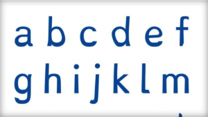
Around 10% of the world’s people have dyslexia, a developmental reading disorder that impairs a person’s ability to read and write. There is evidence that the font of the text has a significant effect on a text’s accessibility for people with dyslexia (Rello, L., & Baeza-Yates, R., 2013). The search for a way to make reading easier for dyslectics lead to the creation of a new type font: Dyslexie. The font Dyslexie was developed especially for dyslectics so that the differences between each character is bigger, easier to recognize, and less likely to be confused with another (Shallow, 2014).
How it works
The Dyslexie font comes with some interesting modifications to letters.
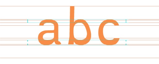
Letters now have what is called a heavy bottom. Dyslexie acknowledges that letters can be viewed as 3D objects. If letters are 3D objects, then gravity applies. Dyslexie “weighs down” the bottom of each letter by making the bottom section bolder, preventing it from tipping upside down.
There are also new slanted parts to the letters. This means that characters which look quite similar have been adapted by changing the tails, to reduce the similarity and avoid the problem of mirror letters.
Openings in letters that have them are bigger. Supposedly, these airier letters are more distinctive, so it is easier to see them as unique.
By slanting the letters, it prevents them from flipping sides and instead weights them toward one side, so that a lowercase B and D would get less easily confused.
There is also a longer ascender and descender in Dyslexie. Lengthening the ascender and descender of the letters allows the differences of individual letters to be emphasized.
Larger and bolder capital letters and punctuation marks, help prevent sentences from running together.
Creating different heights of letters that are usually twins now have unique features so that they no longer resemble each other entirely. Here, the inner corners of the letters appear at different heights and angles.
The space of the letters is expanded with a higher X-axis, making the characters more distinct.
Dyslexie has larger spacing between letters, with the idea that it makes them less likely to become jumbled together and are more clear to read.
Results
Through these many modifications to the proportions, sizing, and spacing of letters, Dyslexie aims to help people with dyslexia read more fluently with higher comprehension (Boer, n.d.). But does it really achieve these goals? Some scientists who study dyslexia have wondered if the type font actually helps and have performed experiments on the subject.
The conclusion of one study was that reading with the font Dyslexie doesn’t lead to an increase in reading speed. There was however a decrease in the reading errors when dyslectics read words in the Dyslexie font (Leeuw, 2010). This study just indicates a decrease in general reading errors, but what specific kind of errors were made?
Another study used eye-tracking to measure the effect of font type on reading speed. It found that Sans serif, monospaced and roman font styles significantly improved the reading performance over serif, proportional and italic fonts (Rello, L., & Baeza-Yates, R., 2013). This study’s findings go directly against some of the core ideas behind the Dyslexie font. Slanting letters would have similar effects as italics, and changing the lengths and sizes of certain parts of the letters makes them disproportionate.
When asked if the new dyslexia font helped, some dyslexic students shared (Burgess, 2012) :
“It helped at first. I really liked it. I didn’t have to focus as much as with the regular font. The reading was easier. But after a while, the new font got actually annoying for me because I noticed the darker bottoms and I was really glad to go back to the regular reading.”
“My eyes didn’t wander away or got distracted as easily as with the regular font.”
“It really didn’t help me because I am a good reader. Maybe it is more helpful for someone who struggles with reading. Not every dyslexic is the same.”
When asked whether the OpenDyslexic font prevented their brain from turning the letters, the younger students unanimously said that it did help “a little bit”. Others said, “No, not really.”
The older students commented that because of having learned to read with the “Help” method, they didn’t struggle with turning letters anymore, just with skipping words or lines at times. “We struggle with new and complicated words just as much as other non-dyslexic students do. Besides, dyslexia is not just about reading, it is about how our brain works and how we think.”
Not all dyslexics have the same cognitive impairments when it comes to reading. There are lots of different routes and paths towards understanding written words. Some can be damaged and others can be spared. The Dyslexie font can help improve some problems for a portion of the dyslexic population, but it also creates some new cognitive difficulties.
References
Boer, C. (n.d.). Dyslexie Font: The dyslexia font which eases the reading. Retrieved from http://www.dyslexiefont.com/en/
Burgess, T. (2012, October 7). OpenDyslexic: ‘Does the new dyslexia font help?’ Review by teacher and students. Retrieved from http://www.examiner.com/article/opendyslexic-does-the-new-dyslexia-font-help-review-by-teacher-and-students
Leeuw, R. (2010). Special font for dyslexia?.
Rello, L., & Baeza-Yates, R. (2013, October). Good fonts for dyslexia. InProceedings of the 15th International ACM SIGACCESS Conference on Computers and Accessibility (p. 14). ACM.
Shallow, P. (2014, November 11). The font that could help dyslexics read better. Retrieved from http://www.cbsnews.com/news/the-font-that-helps-dyslexics-read-better/
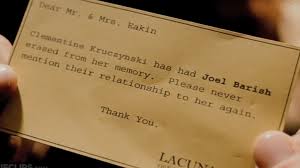
Have you ever wanted to selectively erase your memories of something or someone for good? I bet you have but, with today’s technology, it is not actually possible to choose who and what you can completely erase from your memory. Let’s take the movie, Eternal Sunshine of the Spotless Mind (2004), for instance. This movie is about two lovers, Clementine and Joel, who went through a painful break up and decided to have procedures done that would erase their memories of each other. Joel asked the doctor whether getting the procedure done would cause any brain damage and the doctor responded, “technically speaking, the procedure is brain damage.” Click below to watch a short clip of the movie:
Eternal Sunshine demonstrates a vivid understanding of how the brain forms memories, specifically those that are tied to intense emotional experiences. The movie plays on this idea that the brain stores emotional memories differently from memories that aren’t so emotional; therefore, we can conclude that traumatic experiences are tied to these emotional memories. Traumatic memories are stored in two parts of the brain: the hippocampus and the amygdala. The hippocampus does the job of converting short-term memories into long-term memories, while the amygdala is ultimately the center of emotions within the brain. An important fact to think about regarding traumatic memories is that if a person has hippocampal damage, they will no longer be able to form long-term memories; however, they might still be able to form subconscious traumatic memories if their amygdala is fully functioning. In the movie, Eternal Sunshine, Clementine constantly finds herself subconsciously thinking about emotional memories from her past even though those memories were completely erased from her mind in reality.
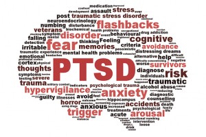
Post-traumatic Stress Disorder
Post-traumatic Stress Disorder, more commonly known as PTSD, is a mental health condition that is caused by a traumatizing experience in the past that may cause flashbacks, nightmares, and uncontrollable thoughts after the event has occurred. In a recent study, however, psychiatrists McGill and Harvard tested the use of an amnesia drug called propranolol, which lends itself to the suppression of emotional factors in memories for trauma victims. Ultimately, what this does is allows people with PTSD to still retain full details of their memories, but the drug lessens their chances of feeling overwhelmed by those memories.
Neuroscientists and psychologists have also been able to develop more theories into the neural mechanisms of how memories are made and stores by means of newer, and better, imaging technology. They have found physiological interventions such as electrical currents and pharmacology that have proven to destabilize fearful memories in victims of PTSD. These victims are encouraged to create newer and safer mental associations to the same sensory cues having to do with whatever memory is causing them anxiety. Long-term memories have enough plasticity, when recalled, to be updated. The victim “updates” their memory by placing themselves in a non-threatening environment simulating their fears, with hopes that they will be able to gain control of them. This activity requires neural communication between the hippocampus and the amygdala. The hippocampus, in this case, cues the prefrontal cortex of any changed conditions in the brain while the amygdala produces a conditioned fear response.
Selectively Restoring Memories
A recent study that comes from the UCLA, Mary S. Easton Center for Alzheimer’s Disease Research and the Buck Institute for Research on Aging have provided evidence that patients with memory loss may be able to reverse their losses. Among patients that were tested were those who suffered from Alzheimer’s Disease (AD), Amnestic Mild Cognitive Impairment (aMCI), and Subjective Cognitive Impairment (SCI). These patients showed a sustained improvement in their memory 3-6 months after the program started. Researchers were able to obtain such results from a complex, 36-point therapeutic program that involved changes in the patient’s diet, exercise, amount of sleep, brain stimulation, specific medicines and vitamins, and many other factors that would affect brain chemistry.
Dale Bredesen, Professor of Neurology and Director of the Easton Center at UCLA, furthered his research to get a better understanding the foundation of Alzheimer’s Disease and why it has had the least amount of success over the past decade in clinical trials aimed at stopping or slowing down the progression of the disease. According to Bredesen, Alzheimer’s Disease comes from an imbalance in nerve cell signaling, which means that the nerve connections are disturbed, therefore causing a loss of memory. Bredesen proposed a multi-component system that is personalized to each patient separately in order to determine what is affecting the plasticity signaling network of the brain of patients with Alzheimer’s Disease instead of testing a drug that only targets a single network in the brain. The downfall to this experiment was its complexity in that it was designed for the patients to follow strict guidelines in changes to their lifestyle- the patients were unable to stick to the protocol.
eRATicate that Memory
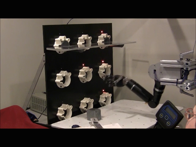
In 2014, researchers at the University of California, San Diego School of Medicine tested on rats to determine whether they could destroy a fearful memory from the animal’s past then reactivate that same memory. This memory reactivation process was done by stimulating nerves in the brain that are known to weaken and strengthen nerve cell connections, otherwise known as synapses. The scientists genetically modified nerve cells in the rat brains to make them sensitive to light, then stimulated these nerve cells as they delivered an electric shock to the rat’s foot. Repeated trials caused the rats to associate the administered shocks with pain, therefore displaying fear behaviors as a result of these shocks.
Following the fearful responses from the rats, the scientists proceeded to weaken the stimulation of the same nerves, which caused a reduction in the rat’s fearful behavior, suggesting that the pain associated with the memory being stimulated was no longer present. Following the same procedures, the scientists attempted to reactivate the rat’s fearful memory by re-stimulating the same genetically modified nerves and the rats subsequently reacted with fearful behavior. Ultimately, the scientists caused the rats to have fear for something, took that fear away, then created that fear again by stimulating selective nerves in the rat’s brain at frequencies that would either strengthen or weaken the synapses.
Even if you have, go find somewhere comfortable and quiet to sit. Take a deep breath and clear your mind. Watch the video below and follow the instructions.
How are you feeling now? Calmer? More relaxed? While meditation used to be primarily for spiritual purposes, it is now used as a form of relaxation and stress reduction. It’s been shown to help emotional well-being and overall health. Research has shown that meditation has the potential to increase your ability to pay attention.
What is attentional blink?
Attentional blink occurs when we don’t notice something shortly after it occurs after something else. To test this in the lab, a series of objects are shown in rapid succession. A subject is asked to identify two target objects out of the series. If the second target object is shown soon after the first, the subject will be “blind” to it. The sensation of not being able to see the second object is what is known as attentional blink.
What causes attentional blink?
There are numerous theories as to what causes attentional blink:
Inhibition Theory
Confusion between the two target images occurs during the target identification process. This causes a gap in attention.
Interference Theory
Other items in the series of target images interfere and compete for limited mental resources.
Attentional Capacity Theory
The process of identifying first target image is too strenuous, causing the brain to not have enough resources to identify the second image in time.
Two-Stage Processing Theory
Processing takes two stages: noticing the targets, and processing the items. Attentional blink occurs when the second target appears while the first target is being processed.
Diagram from the Slagter et. al. (2007) article showing the design of the task used to test attentional blink.
The effect of meditation
Slagter et. al. (2007) conducted an experiment to explore a possible connection between the effects of meditation and performance on attentional blink tasks. The subjects were divided into two groups. The novice group, after being given a 1-hour meditation instruction session, meditated for 20 minutes each day over the course of a week. The practitioner group attended a three-month long retreat where they meditated for 10-12 hours each day. After this, both groups were subjected to attentional blink tests and had their scalp-related brain potentials measured during the test. The study found that the practitioners had far fewer occurrences of attentional blink than the novice subjects, and they managed to allocate their limited amount of attentional resources more evenly between the first and second targets. These findings suggests that meditation has the ability to decrease the likelihood of experiencing attentional blink and to improve resource allocation in the brain.
Why should I care?
Why is attentional blink so important? Coursework from MIT poses the following scenario:
“Let’s say a crime was committed, and they pull the people who witnessed it and ask, ‘How many gunshots did you hear?’ Remember, if you hear one gunshot, you may panic and not notice the next couple of gunshots. And different people will give different reports of how many criminals there were, how many gunshots they heard, and so on. This is a problem in situations where we need to count or keep track of every occurrence of something.”
Attentional blink should be considered in any scenario where a split-second decision needs to be made, such as while driving. Meditation might improve your awareness of your surroundings and make sure that you take in all of the available information before making a decision.
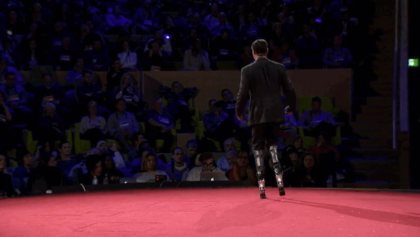
Losing a limb can be a traumatic experience. Hugh Herr experienced this first hand when he lost both of his legs after getting trapped for days in a blizzard while hiking. Today, he can walk, jump, climb, and dance–all thanks to the technology his biomechatronics lab at MIT developed.
Hugh Herr is the head of the biomechatronics research group at the MIT Media Lab, and is an active user of the lab’s inventions, having lost both of his legs. His prosthetic limbs receive input from his residual limbs through electrodes that measure the electric pulses of his muscles. Researchers at Herr’s lab design their prosthetics to mimic how the limbs work normally, so that the person using them can move the limb in a natural way, experiencing no impairment doing day-to-day tasks. Herr demonstrated his bionic legs during his TED talk, which also featured another dancer using a prosthetic.
Hugh Herr demonstrating his dancing skills at his TED Talk.
Herr refuses to think of the human body as “broken” — it is the technology that is broken and inadequate, and his work in the field aims to fix this.
The prosthetic has certain algorithms (such as walking down the stairs) that get activated if the user activates certain muscles in specific ways. This way, the person can transition from walking to doing another action easily. These actions may be tailored for the person depending on their body traits and preferences, allowing individuals to enjoy doing tasks they could do prior to their injuries.
Cortical Prosthetics
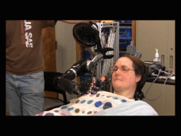
Reading muscle input is not the only way to control prosthetic limbs. Researchers at Johns Hopkins and University of Pittsburgh have managed to monitor activity in the motor cortex of amputees in order to allow them to use prosthetic limbs with ease and fluidity.
This patient is feeding herself a chocolate bar through activations in her motor cortex. Source
This technology requires microelectrodes to be implanted in the brain permanently, and an algorithm extracts the intended movement based on the firing rate in the monitored areas. The patient trains with the device for a number of weeks in order to achieve good control over the limb but is able to freely move it around after a single day of training.
The researchers at U Pitt explain that “when the subject recognizes that her movements are ‘off,’ she can learn to correct them by modifying the patterns of her own neural activity that is being recorded by the electrode arrays.” This means that the patient can learn to use the limb better as they get more experience with it–the researchers don’t necessarily need to tailor every detail for the patient.
This prosthetic arm, controlled through the motor cortex, allows for seven degrees of freedom (three-dimensional translation, three-dimensional orientation, one-dimensional grasping). Early trials were conducted using monkeys, and mostly consisted of making the monkey feed itself. Researchers observed what they call “embodied control” when a monkey started to lick its fingers during a trial, outside of the task requirements. The teams working on this technology have received tremendous amounts of funding from DARPA, and will be developing the product further.
The same patient grabbing a cube to place it on a shelf. Source
Targeted Muscle Reinnervation
There are even more ways of allowing a person to control a prosthetic limb. There is a surgical method called Targeted Muscle Reinnervation (TMR) that uses electromyography (EMG) to detect nerve signals from muscle groups near the site of amputation, and uses these signals with a sophisticated prosthetic to give the patient control over the prosthetic arm. This allows for the person to do various motor movements with his/her arm at the same time, which is not possible with other methods. For example, the person can twist their wrist while opening or closing their arm at the same time. A small caveat is that this method is developed only for arm prosthetics, and will not work for individuals who are unable to use their muscles near the area of amputation, because EMG requires those muscles to be functional. However, this method is widely available and is offered through many hospitals around the US.
The future of prosthetic limbs is bright–when Hugh Herr had his accident 30 years ago, he couldn’t dream of being able to move his prosthetic legs at will, but today he is using one of the most sophisticated prosthetics available, developed through his own research. Prosthetics are becoming less invasive and easier to control, and soon enough, it will be hard to distinguish prosthetics from real limbs.
Sources:
Au, S., Berniker, M., & Herr, H. (2008). Powered ankle-foot prosthesis to assist level-ground and stair-descent gaits. Neural Networks, 21(4), 654-666.
Collinger, J. L., Wodlinger, B., Downey, J. E., Wang, W., Tyler-Kabara, E. C., Weber, D. J., … & Schwartz, A. B. (2013). High-performance neuroprosthetic control by an individual with tetraplegia. The Lancet, 381(9866), 557-564.
Kuiken, T., Miller, L., Lipschutz, R. D., Stubblefield, K., & Dumanian, G. (2005). Prosthetic command signals following targeted hyper-reinnervation nerve transfer surgery. In Engineering in Medicine and Biology Society, 2005. IEEE-EMBS 2005. 27th Annual International Conference of the (pp. 7652-7655). IEEE.
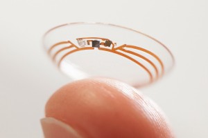
Imagine approaching the intersection of a busy road, but you cannot see a thing. The world is dark, and you must depend on your other senses to get yourself across this road alive. Would you cross? Google’s new smart contact lenses say yes.
A small sensor and a wireless chip placed between two soft contact lenses
The Idea
The original idea behind the development of these smart lenses was to help persons with diabetes. Diabetics deal with daily emotional and physical struggles of having to constantly check their glucose levels, prick their fingers, and get insulin shots. In order to try and relieve this stress, Google decided to insert a tiny chip on a contact lens, which is able to measure the glucose content in a person’s tears. If the glucose levels were to suddenly drop or rise, the lens would alert the person through an indicator such as a blinking light.
Taking this idea a step further, Google designed lenses that are applicable to those who experience a loss of sight. The wearer has the ability to control the lenses with eye gestures, such as blinking, in order to take a picture.
Helping the Visually Impaired
This technology can be used to detect light, colors, patterns, objects, and motion through images that are gathered through the lens. Combined with face recognition software, it would allow a blind person to identify people they see without even hearing their voices. Even for those with perfectly healthy sight, this is a very powerful tool due to the ability to process data, both locally and remotely, through the lens.
For those who are only partially visually impaired, parts of their visual field that are in their blind spots can be digitally stored. This data could then be displayed at a later point and would allow them to sense more. These images can be easily displayed on a smartphone or tablet.
An example of what a person with a scotoma sees
This technology would be particularly useful for people with conditions such as hemianopia, scotoma, and quadranopia, because although part of the image they see may be blurry due to lesions in their primary visual cortex, a picture of a scene can be rearranged until the patient has the ability to see and fully comprehend the full image.
Let’s return to the intersection example. While approaching this intersection, imagine that the camera in your eye is rapidly processing the nearby field, determining whether there are cars speeding by. Once it detects that the road is clear, the wireless chip sends an alert to the your smartphone to let you know that it is now safe to pass. This technology communicates visual information through auditory channels, allowing individuals to use senses other than sight alone.
More specifically, these lenses are applicable to those with presbyopia (no longer being able to view objects closely, which results from a loss of elasticity in the lens of the eye). Research suggests that people with presbyopia should have the ability to read or see close up using the Google contact lenses because they “provide accommodative vision correction to help restore the eye’s natural autofocus on near objects in the form of an accommodative contact lens or intraocular lens as part of the refractive cataract treatment.”
Future Directions
There are endless possibilities for how this smart lens can help people live more comfortably, even without the full use of their vision. Unfortunately, there are a few caveats to this invention. People with physical damage to their eyes may not be able to wear contact lenses, making this device more applicable to those who lost their sight due to brain damage, not direct eye damage. Also, this technology is more desirable for discrete face recognition and documentation purposes, perhaps to be used by law enforcement, rather than to help the visually impaired. We don’t yet know what will come out of this work-in-progress, but it is certain to enhance the capabilities of the eye.
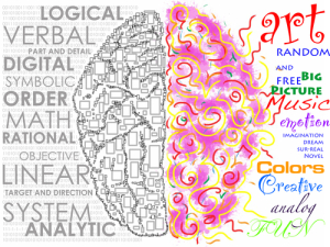
Hemispheric dominance is the idea that both hemispheres of the brain are designed to perform specialized, but different tasks. Both sides of the brain control and receive sensory input from one another, which is known as contralateral control. The performance of tasks may also be referred to as “lateralization.” The theory of hemispheric dominance, however, has been oversimplified for many years due to people confusing the actual science with personality traits.
For example, many people associate the left hemisphere with mathematical skills and logistics, whereas the right hemisphere is known for artistic and imaginative skills. But why is that? It is true that both sides of the brain carry out specific functions, but they should not be categorized as two separate ways of thinking. Many processes are carried out in both sides of the brain. For example, the left side of the brain is dominant when processing speech, even though speech is processed using both sides of the brain.
All of the very complex behaviors and functions of the brain are shared with different regions on both sides of the brain; therefore, this idea of being right-brained or left-brained is a myth, perpetuated in part by some misinformed “scientific articles”.
—–
On their website, UCMAS Math Schools make the claim that, “By learning abacus through the systematic training approach at UCMAS, children can fully realize their potential by activating both sides of their brain. By consciously using the right side of our brain, we can be more creative.” It is not possible to “consciously” use a specific half of our brain, and our hemispheres do not need to be manually activated. This school is interpreting the concept of hemispheric dominance in a false way to create pseudo-scientific support of their use of abacuses.
UCMAS’ website shows that they are a tutoring program, but they fail to cite any sources in their article on left and right brains. Also, once you click on “More Info on Brain Development”, it brings you to a page which states the following: “Recent studies have proven that the abacus method of mental calculation is extremely effective in activating the right brain. According to the research, the learning and thinking process is enhanced when both sides of the brain participate in a balanced manner.” Of course, none of the studies mentioned in this section are cited. It becomes quite clear that the article is a marketing attempt for getting parents to sign their children up for classes.
—–
Iain McGilchrist, in an article written for the Wall Street Journal, talks of the two hemispheres of the brain as two separate worlds, and says that one hemisphere can come to dominate individuals and even whole cultures. He claims that, “Without the right hemisphere, we are socially and emotionally insensitive, and have an impaired understanding of beauty, art and religion”. This implies that we could actually live without one half of our brains. He goes on, explaining how life is without the left hemisphere and how throughout the ages, certain cultural changes have allowed one hemisphere or another to dominate. He is particularly concerned about Western culture, saying that the result of over 2500 years of mental battles is “an ever greater reliance on the left hemisphere”.
The Wall Street Journal article seems pretty credible at first; after all, it comes from a major publication. But on a closer look, the article comes from their Life and Style section, rather than from their Science section. And the information at the end of the article about the author, he wrote it to promote his new book.
—–
Anandi, the “sleep guru” claims on her website that the left hemisphere is masculine, bearing qualities “such as logic and reason”, while the right hemisphere is feminine, acting as our source of “intuition, creativity and spontaneity”. Which not only enforces wrong information about the brain, but also enforces incorrect gender roles. She then implies that the difference between the openness of our nostrils is connected to the balance between the the hemispheres.
The sleep guru site is a personal blog/webpage devoted to helping its owner, Anandi, gain new clients. Anandi helpfully lists her credentials on her “Who is the sleep guru?” page. While I am sure that her qualifications make her an excellent meditator and yoga instructor, she has no experience in neuroscience and is therefore not a credible source on hemispheric dominance.
——
There is a lot of misleading information on the internet, which can make it hard to find accurate information about hemispheric dominance online. The first result that pops up when trying to research hemispheric dominance is a link to a quiz which claims to help you determine which hemisphere is most dominant, are you right brain or left brain quiz. Pop culture has adapted hemispheric dominance and turned it into a concept with little scientific basis. It’s important when researching hemispheric dominance, or any other topic, to make sure that the information is coming from a credible source.
 Letters now have what is called a heavy bottom. Dyslexie acknowledges that letters can be viewed as 3D objects. If letters are 3D objects, then gravity applies. Dyslexie “weighs down” the bottom of each letter by making the bottom section bolder, preventing it from tipping upside down.
 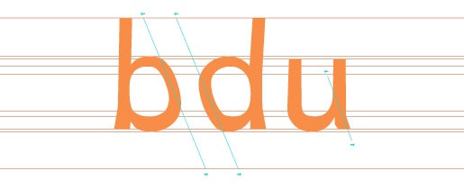 There are also new slanted parts to the letters. This means that characters which look quite similar have been adapted by changing the tails, to reduce the similarity and avoid the problem of mirror letters.
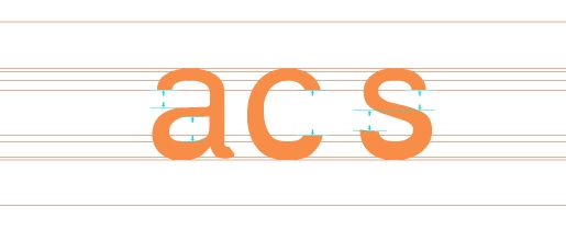 Openings in letters that have them are bigger. Supposedly, these airier letters are more distinctive, so it is easier to see them as unique.
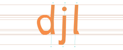 By slanting the letters, it prevents them from flipping sides and instead weights them toward one side, so that a lowercase B and D would get less easily confused.
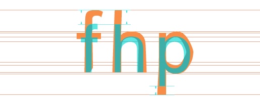 There is also a longer ascender and descender in Dyslexie. Lengthening the ascender and descender of the letters allows the differences of individual letters to be emphasized.
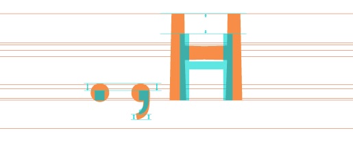 Larger and bolder capital letters and punctuation marks, help prevent sentences from running together.
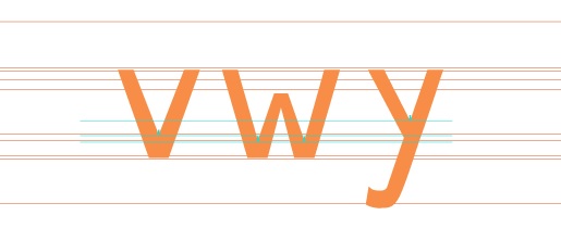 Creating different heights of letters that are usually twins now have unique features so that they no longer resemble each other entirely. Here, the inner corners of the letters appear at different heights and angles.
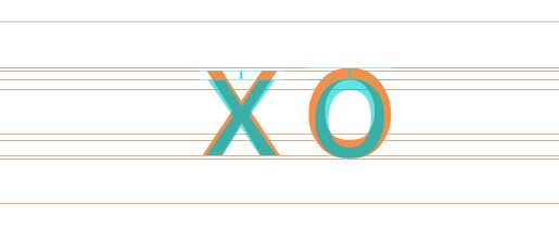 The space of the letters is expanded with a higher X-axis, making the characters more distinct.
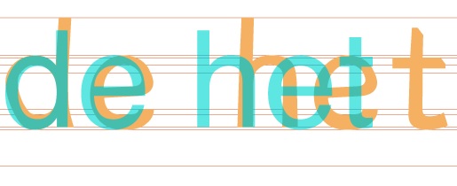 Dyslexie has larger spacing between letters, with the idea that it makes them less likely to become jumbled together and are more clear to read.
 Not all dyslexics have the same cognitive impairments when it comes to reading. There are lots of different routes and paths towards understanding written words. Some can be damaged and others can be spared. The Dyslexie font can help improve some problems for a portion of the dyslexic population, but it also creates some new cognitive difficulties.

 ich lends itself to the suppression of emotional factors in memories for trauma victims. Ultimately, what this does is allows people with PTSD to still retain full details of their memories, but the drug lessens their chances of feeling overwhelmed by those memories.
 SCI). These patients showed a sustained improvement in their memory 3-6 months after the program started. Researc
 n as synapses. The scientists genetically modified nerve cells in the rat brains to make them sensitive to light, then stimulated these nerve cells as they delivered an electric shock to the rat’s foot. Repeated trials caused the rats to associate the administered shocks with pain, therefore displaying fear behaviors as a result of these shocks.
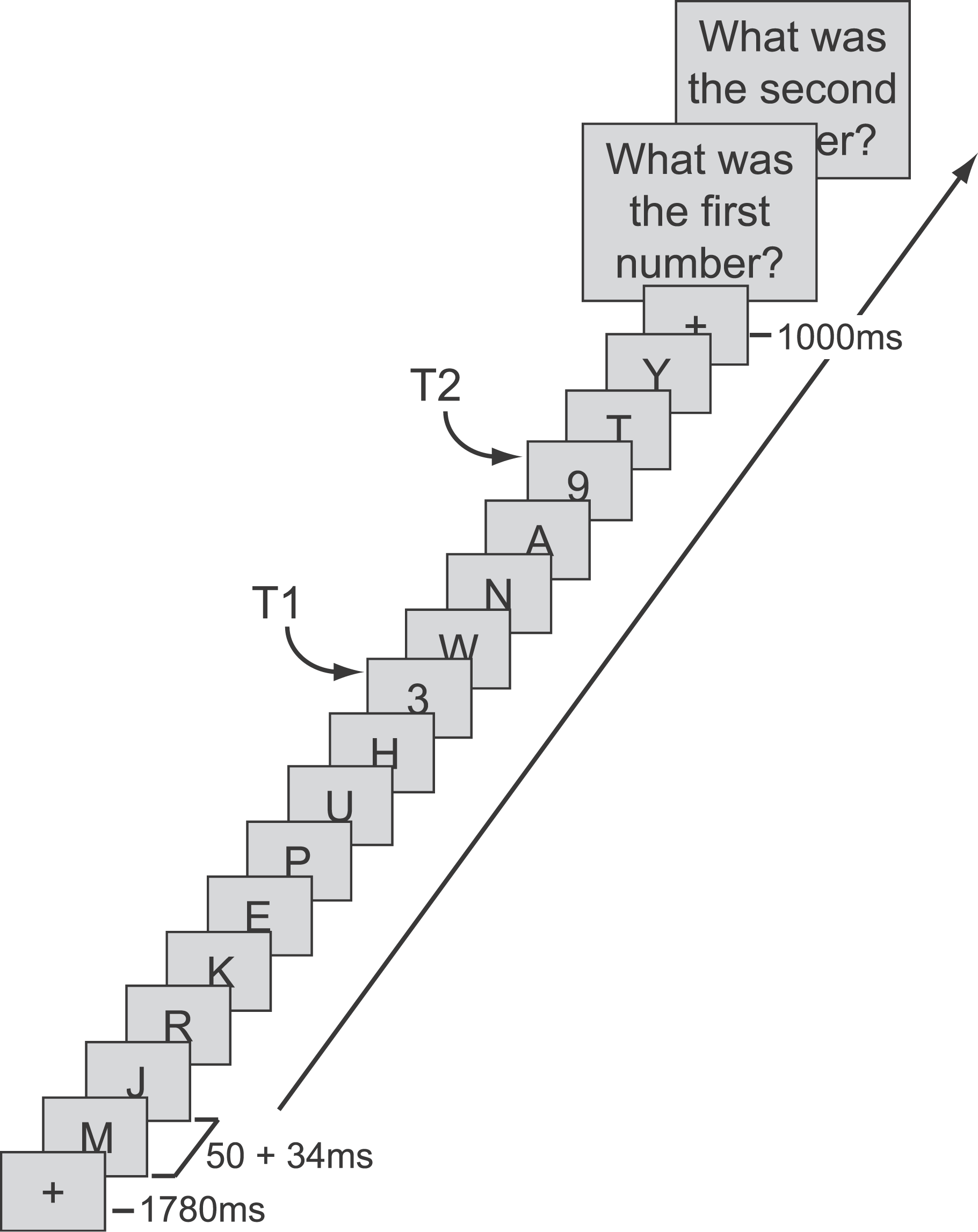

